


Lumbar tumoris one of the most common diseases of the spine.
It is characterized by deformation of the cartilage tissue of the vertebra.
The spine remains flexible and mobile as long as the vertebrae are healthy.
If the condition gets worse, the discs lose elasticity and start to dry.
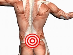
As a result, the patient suffered severe pain in the lumbar region.
What is it?
As degenerative-dystrophic changes appear in the tissues of the vertebra, they begin to deflate. If the vertebrae are located in the affected lumbar region, then lumbar spondylolisthesis is diagnosed.
Clinical presentation
With osteonecrosis, the cartilage tissue of the vertebra begins to lose moisture, and the elasticity of the disc becomes less resilient. Over time, the height between the vertebrae decreases. Under the increasing load, the lint starts to crack, the disc bulges outwards.This leads to pinched nerve ends and pain.
In the absence of proper treatment, bone necrosis will progress. The discs harden, their cushioning properties deteriorate. Development occurs on the bone tissue of the vertebra, compressing the nerve ends. Therefore, the patient suffers from constant pain.
Level and category
Experts distinguish 4 levels of spinal tumors:
- Discardment begins, symptoms of mild illness, a burning sensation, and tingling appear only after exertion. Patients talk about the appearance of dull back pain, sometimes they spread down the buttocks.
- The distance between the vertebrae gradually decreases, and the annular vertebrae begin to collapse. Squeezed discs, beyond physiological boundaries, put pressure on nerve roots. The patient complains of obvious pain, during walking given in the buttocks, thighs and legs. In addition to pain, there may be a feeling of hot, cold.
- The loops are destroyed, and on examination the patient discovers a herniated disc. Persistent pain, regardless of the load.
- Bone growing from vertebrae can be seen. Cartilage atrophy, patients have difficulty moving. As a result, the lumbar spine loses the ability to move and flexibility, the patient is disabled.
| Bone tumors are also categorized by disease course: | |
|
|
The following experts distinguish bone necrosis stage:
- released;
- aggravated;
- relieved; Stable
- .
Treatment is chosen depending on the stage of the disease, the nature of the malformation and the severity of symptoms of the disease.
ICD 10 Code
According to the international classification of the disease, spinal necrosis disease has been coded M42. Separately, there are juvenile (M42. 0), mature (M42. 1) and unspecified (M42. 9) osteonecrosis.
popularity and importance
The lumbar region is more susceptible to bone necrosis than other areas of the spine. This is because the load on this area increases, as it has to support the body weight. With a weak mechanical corset, the condition of the discs starts to deteriorate rapidly, they are destroyed.
Normally, people who have passed the 30-year markhave necrosis of the lumbar spine. Although it can be seen in younger patients. Nearly 80% of patients who see a doctor with pain in the lumbar region are diagnosed with bone necrosis.
Examination of patients over 40 years of age shows that most of them have characteristic changes in the disc. But in the absence of clinical manifestations, a person is not considered ill.
In the absence of adequate treatment, the disease will progress. In neglected forms, it leads to the disability of the patient.
Risk Factors and Causes
Often, representatives of such professions encounter osteonecrosis: programmers, office workers, builders, drivers, waiters, and drivers.

Risk factors, when there is an increased likelihood of developing osteonecrosis, include:
- overweight;
- unhealthy diet;
- problems with posture; genetic predisposition
- ;
- lack of sleep;
- is often stressful;
- continuously cool down;
- needs to be in an uncomfortable position for a long time;
- low physical activity.
Reasons for the development of lumbar spinal necrosis include:
- the body's natural aging process;
- metabolic problems;
- back injury;
- return issues;
- flat feet;
- intense physical activity, eg weightlifting;
- problems with spinal joints (rheumatoid arthritis);
- endocrine diseases;
- problems with the digestive and cardiovascular system.
Some experts believe that the tendency to develop osteonecrosis is passed down at the genetic level.
Consequences
A change in the cartilage tissue between the vertebraeleads to degeneration of the fibrosis and the appearance of a hernia. Patient begins to complain of severe pain in the lumbar region, spreading down to the glutes, thighs, and lower legs. But this is not the only possible complication of osteonecrosis.
Prolonged irritation of the spinal cord leads to inflammation.Patients with lumbar sciatica.
With osteonecrosissciatica may develop(sciatica). The disease leads to severe pain, numbness in the back and legs. The patient begins to walk, leaning to one side. This causes further curvature of the spine and further destruction of the disc.
Osteoporosis causing instability of the vertebrae. The lumbar region, under the action of body weight, begins to move from the sacrum. In women, such an instability leads to the appearance of problems with internal organs (uterus, ovaries, affected appendages), in men - with the possibility.
When the discs are destroyed, blood supply to the spinal cord is interrupted, displacement of the vertebrae leads to compression of the myelopathies.
Equina Cauda syndrome is considered the most dangerous complication. It includes the fact that the nerve roots are affected. In some serious cases, osteonecrosis causes paralysis of the lower extremities or paralysis of both legs.
It is possible to prevent the development of negative consequences if, when symptoms first appear, consult a doctor and do not ignore the need for treatment.
Symptoms
Osteochondrosis does not appear immediately. In the early stages, the patient does not feel any pain or discomfort. Usually the complaints appear once the disease progresses to stage 2.
Key symptoms of lumbar necrosis include:
- low back pain worsens as the disease progresses;
- decreased mobility: problems when trying to bend over, turn around, feeling when changing body position is described by the patient as "electric shock", many pain spread downfoot;
- changes in the sensitivity of the extremities, appears on the basis of nerve root damage, in the affected area there is a feeling of burning, numbness, ants, tingling;
- muscle weakness, lack of tendon reflex;
- reduces local temperature;
- increased sweating;
- pallor, dry skin in problem area;
- urination disorders, sexual dysfunction (in severe bone necrosis).
Some patients have spasm of the arteries in their legs. But symptoms manifest only in the form of acute bone necrosis. Exacerbations may start suddenly with hypothermia, awkward movements or after intense physical activity.
Which doctors are treating?
If you have back pain you shouldsee an orthopedic and neurologist. Examining the patient's neurological condition, examining how the spine functions. Doctors also evaluate the condition of the back and butt muscles.
For experienced specialists, one visit is sufficient for a preliminary diagnosis. But to confirm it, the patient is sent to diagnose the hardware.
Diagnostic method
The simplest and most accessible method to detect bone tumors isx-ray. But to get more accurate pictures, the computer or magnetic resonance imaging is prescribed.
MRIhelps check spinal condition as accurately as possible. Indeed, in the process, layered photos of the problem area are taken.
Treatment

Treatment strategies are chosen by the doctor depending on the patient's condition, the stage of bone necrosis and the clinical manifestations of the disease.
A doctor can prescribe:
- drug therapy, nonsteroidal anti-inflammatory drugs, hormonal drugs, pain relievers of choice;
- blockers, pain relievers, hormonal drugs injected into the affected area or problematic surrounding muscles, almost immediately reduces inflammation andeliminate pain;
- manual therapy, massage, physical therapy, is recommended after stopping the acute phase of the disease, with the help of physical therapy, you can increaseincrease the effectiveness of drug therapy;
- medical gymnastics; Acupuncture
- .
Required in advanced cases. Surgical interventions are indicated in situations where conservative treatment does not produce the expected results.
Conclusion
With progression ofdystrophic degenerative changes in the cartilaginous tissues of the lumbar spine, osteonecrosis disease is diagnosed. In severe form, this disease can not only lead to the appearance of intense pain continuously but also cause paralysis, paralysis of the lower extremities.
- You can suspect the development of osteonecrosis when lower back pain is present. With the progression of the disease, the pain increases significantly, the lower back gradually loses its ability to move.
- Depending on the degree of destruction of thedisc, the disease has 4 stages.
- This diagnosis usually applies to people after age 30. Nearly 80% of patients who see a doctor for back pain are diagnosed with osteonecrosis.
- People with inactive lifestyles are susceptible to bone necrosis, which is in an unnatural position for long periods of time, often with physical overload.
- The main symptoms of osteonecrosis are pain and impaired lower back mobility.
- Due to destruction of the lumbar spine disc, the patient developed leg defects.
- If left untreated, pain increases,sciatica, spondylolisthesis, spinal cord compression may develop. In advanced cases, it paralyzes the lower extremities.
- In case of painconsult a neurologist and orthopedist. Patients are sent for x-rays, computed tomography or magnetic resonance imaging.
- Depending on the medical condition,is prescribed medication, blockade, massage, manual therapy, physical therapy, physical therapy or surgical exercises.