


The back muscles were over-stretched causing a lot of discomfort and pain. Osteochondrosis, which causes a structural violation of the vertebra and the disc, leads to severe compression of the nerve ends. Usually, the pathology is accompanied by a decrease in blood circulation, causing a disruption in the nutrition of the brain and internal organs.
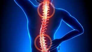
Osteochondrosis - what is it?

Osteochondrosis is a recurrent disease that occurs in a chronic form and is accompanied by destruction of the vertebrae along with the intervertebral discs. Their tissues are disturbed, resulting in a decrease in their elasticity, resulting in a change in shape. There is a gradual decrease in disk space. This causes an instability of the spine in the developed regions of the pathology.
Pathological tissue destruction processes occur on the background of pinched nerve ends, which are drained from the area of the spinal cord. As a result, the muscles of the back are always in tension. In such situations, the patient complains of back pain and other symptoms.
Based on the peculiarities of the location of the structures of the spine, covered by degenerative changes, the types of the cervical, thoracic spine and spine of the pathological process are distinguished. The main symptom of the development of osteonecrosis is pain, the intensity and severity often increased with exertion.
Can also freeze in motion. In addition, the clinical picture is characterized by the presence of signs of the type of vertebrae - headache, changes in blood pressure, impaired visual function, hearing, etc. v.
Development Mechanism
The development of osteonecrosis is related to the fact that the medullary nucleus begins to lose hydrophilic properties. This semi-liquid structure contains connective tissue fibers and chondroitin, a viscous substance. During the development and growth of the human body, the processes of reducing the vascular wall in the disc are active. The nutrients are supplied in a diffuse manner, expressed in spontaneous stability of the concentration. This feature becomes the reason it is difficult to completely restore cartilage that has been damaged or under great pressure on the spine.
Pathological abnormalities become more prominent due to a violation of the human hormonal background and nutrition. Cartilage tissue begins to lack nutrients needed for its normal development. Hence, the confusion appears in the form:
- decrease in strength and elasticity;
- changes consistency parameters and configuration properties.
In the context of a flat disc, radial cracks form in the annular fibers. As a result, the spacing between the discs is reduced and the joints begin to change. Over time, pathological changes cover the types of connective tissue involved in the fibrous rings and ligaments.
When tissues are broken down by the immune system, an increased amount of immunoglobulins are produced. This provokes the development of aseptic inflammatory processes, edema is formed in the area of the joints. They also spread to nearby soft tissues.
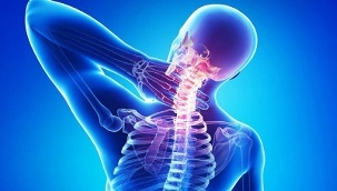
As the synapses are stretched, the discs lose the ability to fix the vertebrae. Such an unstable spinal structure position increases the risk of pinching nerve roots or compressing a vessel. This feature is typical, for example, for cervical necrosis, which is accompanied by severe symptoms.
Cause of the disease
The condition of the disc can worsen with a decrease in skeletal muscle tone in the spine. Due to the mismatched and asymmetrical work of the muscles, destruction of cartilage tissues can occur by long-term preservation of the body's non-physiological position. This offense is the result of wearing a heavy shoulder bag, using a soft mattress and a high pillow.
The process of destroying the disc is accelerated due to the impact of a number of negative factors from the outside and inside the body. These include:
- disorders of the endocrine mechanisms and metabolic disorders;
- pathology of an infectious nature, including in a chronic form;
- damage to the spine in the form of a compression fracture, bruising;
- frequent and prolonged hypothermia;
- systemic diseases and degenerative-dystrophy - gout, psoriasis, rheumatoid arthritis, osteoporosis, osteoarthritis;
- smoking and abusing alcohol, disturbs the state of the vascular system, impaired blood circulation and causes lack of nutrients in cartilage;
- inadequate physical development, posture problems, flat feet - these defects increase the load on the spine, as the amount of wear will not be enough;
- obesity; genetic predisposition
- ;
- is exposed to constant stress.
Symptoms

The main clinical sign of osteonecrosis at any site (cervix, chest, or sphincter) is pain syndrome. When it comes back, the pain penetrates, radiating to the vicinity of the body. Even with a slight movement, it gets stronger. This forces the patient to position himself in a mandatory position to minimize discomfort and pain:
- with cervical bone necrosis, should not rotate one head but the whole body;
- in the presence of thoracic pathology, the patient has difficulty breathing deeply, therefore, to rule out acute chest pain, the patient should try to minimize the depth and frequency of breathing;
- in patients with lumbar disease, difficulty arises when they sit down, stand up, move around, because the nerves of the spinal site are pinched.
Usually, patients complain of a dull, persistent pain and a feeling of stiffness in motion in the morning after waking up. In this case, a differential diagnosis will be required to help rule out the risk of developing myositis due to spondylitis or osteoarthritis.
Soreness and soreness occur due to the compensatory tension of muscle tissue. This is necessary to stabilize the spinal motor area. Persistent mild or moderate pain may be accompanied by significant dilatation of the disc and as a result of aseptic inflammatory changes.
Osteochondrosis of a particular area is characterized by special symptoms:
- With cervical necrosis, pain is felt in the cervical area, in the upper extremities. Look for head swelling and numbness in the fingers. If the disease manifests itself in a serious form, a compression of the vertebral artery may result. In this case, the patient began to complain of a significant decline in health.
- The thoracotomy is manifested by acute pain and back pain, visceral pain syndrome present in the heart, right lower lower and abdomen. The patient complains of numbness, skin paresthesia, dyspnea, and scratching of the vertebrae.
- Patients with lumbar necrosis complain of pain in the back and lower extremities with increased intensity with movement. Most often, disorders in the functioning of the organs of the genitourinary system, male capacity problems, ovarian dysfunction are diagnosed. During remission, the pain may subside. However, the impact of a provoking factor leads to its renewal.
- When manifesting mixed bone necrosis, symptoms may manifest themselves in multiple regions at once. This condition is characterized by a more severe episode.
It is important to remember that vertebrae displacement and the formation of bone cells compress the vertebral artery. It nourishes the brain, providing its cells with an oxygen component. When pressed, food is restricted, and thus the patient has problems with coordination, headache, tinnitus, and arterial hypertension.
The consequences if not treated
The reason for the complex process of osteonecrosis is the relatively rapid formation of herniated masses in the intervertebral discs. Their appearance is associated with the displacement of the vertebral structure in the backward direction. This causes a rupture of the posterior longitudinal ligament of the longitudinal type, resulting in the position of the disc unstable, its individual parts protruding into the area of the spinal canal. A herniated rupture occurs when a disc with a pulp nucleus enters the canal area.
With abnormal manifestations of the vertebral structures, the posterior brain begins to compress, the patient has cervical marrow disease. Symptoms of this condition are related to numbness and weakness in certain muscle groups of the upper and lower extremities. Manifestations of paralysis, muscle atrophy and tendon reflexes. In some cases, there is a problem with the emptying of the bladder, with the intestines.
Disc has a dangerous herniation due to compression of the arteries supplying the spinal cord. The result of this pathology is the formation of ischemic zones where nerve cells are damaged and died. Neurological effects are manifested in malfunction of motor function, decreased sensitivity and sexual disturbances.
Diagnosis

An initial diagnosis is made based on the patient's complaints and symptoms. Experts study the condition of the spine in different positions, suggesting that the patient should rest or move. At the next stage, the patient is sent for laboratory diagnosis, which will help clarify the diagnosis or reject it.
The research methods used include:
- X-ray- provides an examination of the entire spine with an assessment of the condition of the vertebrae, existing disorders in the form of growth, curvature. The specialist will be able to determine the intervals of the disc type, the state of the holes. To accurately determine bone necrosis, localized in the chest area or cervix, a two-stage X-ray examination is performed. In the first stage, the patient lies on his side, and in the second stage, lies directly on his back.
- MRI or CT tomographyprovides highly informative data, helping to study the vertebra in detail without interfering with the form of organ coverthem. Pictures show the nerves and vascular system. MRI helps identify signs of many diseases of the spine and the site of damage. With CT, herniation is visualized, possible deviations in the structure of the spine are determined.
- Laboratory teststo assess the health of the blood and major blood parameters. Allows you to clarify the diagnosis and determine the likelihood of developing concurrent diseases.
In many cases, the examination results, the doctor diagnoses there are some underlying diseases, potentially dangerous complications. For example, we are talking about hernias, bulging eyes, and pulp inflammation. Correct diagnosis of problems helps to effectively treat osteonecrosis. At the same time, the disease itself in the early stages of development is disguised as symptoms of other diseases.
Therapeutic Procedures
Bone tumors undergoing conservative or surgical treatment. The choice depends on the severity of the condition, neglect, the degree of tissue impairment, and the cause.
It is important to remember that osteonecrosis cannot be completely cured as there is no medication to fully restore the disc and vertebrae. The therapeutic effect focuses on inhibiting the destructive process and increasing the duration and stability of remission.
For symptomatic therapy, drugs chondroprotectors are used, based on chondroitin sulfate or glucosamine.
The efficacy of a course of treatment using chondroprotectors has been clinically validated on the basis of long-term trials. If you use these funds for a long period of 3 months, there will be a recovery of the cartilage and other elements of the connection type - the ligament-tendon apparatus, the bursa.

The accumulation of glucosamine and chondroitin in the intervertebral region leads to the manifestation of analgesic, anti-edematous and anti-inflammatory effects. Therefore, there is a real chance to optimize the dosage of NSAIDs, drugs of the glucocorticosteroid group, and muscle relaxants. You can count on reducing your patient's drug load.
The effectiveness of the chondroprotectors is determined by the frequency of their intake. If not, there will be no results. Inefficiency has also been observed in the treatment of grade 3 bone necrosis, which is accompanied by significant cartilage destruction.
The following classes of medications can be used for pain relief:
- Nonsteroidal anti-inflammatory drugshelps to eliminate inflammatory disorders in soft tissues caused by spinal displacement. NSAIDs are effective at relieving pain, swelling, and stiffness.
- Means of the glucocorticosteroid group- often blockade to be used together with anesthetics. They can relieve pain, restore immune mechanisms and provide anti-secretory effects.
- Muscle relaxants.They are effective against muscle spasm due to nerve entrapping. They help relax the muscles of the skeleton and suppress the reflexes of the polyarthritis spine with antispasmodic effects.
- Topical remedies have a warming effect.Irritation of subcutaneous tissue receptors with the activation of the blood stream provided by special gels and ointments. These drugs are characterized by analgesic and anti-edematous effects.
It is possible to eliminate symptoms of spinal allergy, manifested by the localization of the pathology in the area of the cervix or chest, with the help of medical devices to trigger blood flow. Nootropics and drugs to improve microcirculation are also prescribed. In some cases, antidepressants, as well as anticonvulsants may be needed.
During the treatment of osteonecrosis, physical therapy is also used. UHF therapy, magnetotherapy, laser therapy, reflexology, massage, exercise therapy, hirudotherapy, as well as swimming and yoga can be prescribed. If conservative therapy is ineffective, surgery is performed using microscopic resection, disc puncture, laser reconstruction, or implant replacement.