


Herniated discs commonly present in age-related osteonecrosis due to annular fibrosis are dry and brittle. But this is just one of the risk factors. Others include:
- Lumbar strain due to being overweight.
- Weakness of the mechanical apparatus.
- Genetic.
- A sedentary lifestyle and the resulting constant compression of vertebral structures.
- Smoke.
- Great physical activity.
According to medical statistics, this disease occurs many times more often in men than in women.
What are the most common causes of the disease:
- Injury from a road accident or fall.
- Lifting heavy objects with incorrect load distribution.
- Scoliosis or curvature of the spine, leading to increased stress on certain areas of the spine.
- Hip dysplasia.
- Chronic diseases, including tuberculosis of the spine, cancer, syphilis.
- Metabolic disorders (inherited and acquired).
All these factors cause the cartilage and bones of the spine to wear down and become weaker. And this is the main cause of herniated disc disease.
Stages of disease development
Without proper treatment, the disease will progress to worse and the condition of damaged spinal discs will become more and more severe. There are four stages of disease development:
- The fall. The disc has moved slightly, no more than two millimeters. The nucleus pulposus does not protrude beyond the vertebral body.
- Exophthalmisis. The edge of the disc extended beyond the vertebral body to 1. 5 mm, but no nuclear displacement was observed.
- Extrusion. The nucleus protrudes outside the vertebral body.
- Sequence. The nucleus actually falls out and hangs on the vertebrae in the form of drops. At this stage, there is a risk of rupture of the fibrous rings and leakage of secretions.
In the early stages of the disease, the patient hardly worries about anything, there are occasional back pain but quickly passes. With the development of the disease, the state of health also deteriorates, the painful symptoms are more and more alarming. If not diagnosed on time and not started treatment, the consequences can be: paralysis of the legs and serious disorders of the nervous system.
How does a unilateral hernia manifest?
A herniated disc can manifest itself with the following symptoms:
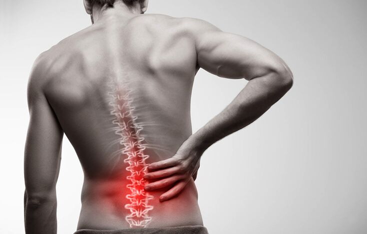
- Pain in the lumbar region.
- Pain when walking, spreading to the thigh area.
- Numbness in the feet, fingers, and areas of the lower legs and thighs.
- Feeling of heaviness in the legs.
- Motion stiffness.
In order not to delay in seeking medical help, it is necessary to analyze the symptoms of the disease more carefully. They can be divided into three groups.
Pain syndrome
Pain with a lumbar herniation is a major symptom. Even in the early stages, the damaged disc area is also painful, especially after trauma. They can increase or decrease, and then arise again. Usually, the sacrum doesn't even hurt, but it does ache, especially with prolonged exertion or sedentary work. If a person lies down on the healthy side and flexes the leg, the pain will completely subside. This condition can last for several months.
With prompt treatment to get medical help, getting rid of the problem becomes easy. You just need to say goodbye to bad habits and do the physical therapy exercises recommended by your doctor.
Every day, the affected area will increase, and the condition of the disc tissues will worsen. The transition to the second degree of the disease is signaled by increased pain. This phenomenon is felt not only in the sacral region, but also covers the entire lower back, radiating to the cervical region, to every muscle of the spine, buttocks, thighs, shins, feet and toes. Discomfort manifested by physical activity, even insignificant - coughing or sneezing.
Vertebral syndrome
Increasing pain in the second stage is accompanied by constant spasms of the muscles in the back. This leads to more discomfort for the patient. The baby cannot freely move, straighten his back, stretch his shoulders. The gait of such a person becomes uncertain, he always leans to the side facing the patient, sagging.
Due to impaired motor coordination, people's quality of life declines. He is unable to perform well at the tasks posed at work, and active rest due to constant pain becomes futile.
Lens syndrome
If the hernia is not taken care of by doctors, the disease will progress to compress the spinal roots, as a result, they die and blood to the tissues of the damaged disc is almost impossible. Symptoms characteristic of severe stages of the disease appear:
- Weak leg muscles. The patient cannot squat, stretch, run or jump. Even climbing stairs was difficult for him.
- Numb the affected area and surrounding areas. The skin becomes emotionless and pale, with goosebumps and tingling sensations. Patients complain of hyperhidrosis in the affected area and legs, or conversely, excessive dryness of the skin.
- Back pain. The patient has low back pain with acute, sharp pain that increases with movement. If left untreated, it leads to destruction of the hip and knee joints.
- The painful leg is significantly thinned, resulting in a disproportionate posture.
- Disruption of pelvic organs. Exacerbation of urological and gynecological diseases, loss of libido, diarrhea, urinary incontinence.
Severe cases of spinal hernia can lead to paralysis, disability, and even death.
Diagnosis of disease
If a person has severe low back pain, he needs to make an appointment with a neurologist. He will conduct an examination with medical tests:
- Identify reflexes from the tendons of the lower extremities.
- Leg lift test.
- Determines sensitivity to heat or cold, pain and vibration on the entire surface of the legs, thighs, buttocks, abdomen and back.
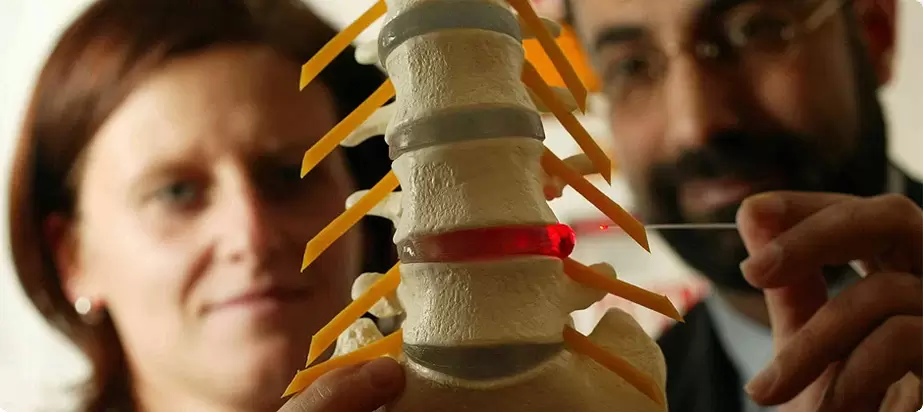
The doctor will then refer the patient for an MRI or CT scan of the lumbar spine. With the help of tomography techniques, a three-dimensional picture of the affected area is obtained. It can be used to determine the location and size of the hernia and the stage of the disease.
If there is a risk of spinal cord injury, electromyography, nerve and spinal cord contrast are additionally indicated. With the help of these studies, the doctor will determine if urgent surgical intervention is necessary.
Herniated disc treatment
Vertebral hernias are treated both surgically and conservatively. The choice of technique depends on the stage of development of the disease, the presence of concomitant diseases and contraindications.
Conservative therapy
The main aim of treatment is to relieve pain and alleviate the patient's condition.
What medications may your doctor prescribe:
- Pain relievers and inflammation. In case of exacerbations - in the form of injections. Once the acute pain subsides (usually three to four days is enough), oral medications with a similar effect will be prescribed.
- Blockade of novocaine with the addition of corticosteroids. A similar method can stop the pain for two weeks at the same time. Usually, an occlusion is done by injecting into different parts of the damaged disc.
- Muscle relaxant of central action. They reduce muscle activity by reducing the pain of cramps.
- Vitamin-mineral complexes concentrate on elements of group B. They provide mild relaxation of muscles, help with tissue regeneration and conduction of nerve impulses.
After the pain syndrome is relieved, the amount of the drug is reduced. Treatment of the disease is through physical therapy and physiotherapy measures.
Physiotherapy treatments are also selected according to the patient's condition. This could be:
- Treat with heat or electric shock.
- Electrophoresis with anti-inflammatory drugs.
- Acupuncture and acupressure.
- Hirudotherapy.
- Mercury acupuncture.
Ordinary massage is allowed only if there is no pain syndrome. A more effective physiotherapeutic treatment is manual therapy with post-isotonic relaxation.
Doctors especially recommend that patients who are smoking should give up smoking.
Nutritional modification is also important, especially for overweight patients. Fatty, salty dishes, sweets and alcohol will have to be excluded from the menu. A diet with lots of vegetables and fermented dairy products will help the body better tolerate the treatment process, as well as remove the heavy weight on the back.
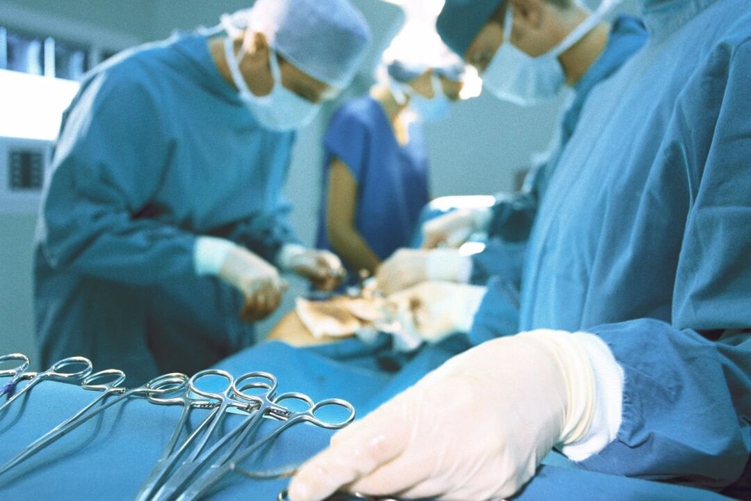
Surgical intervention
Conservative treatment usually lasts about two months. If it does not yield the desired results, a decision is made to change treatment tactics or perform surgery. The latter drug is prescribed for severe pain, loss of sensitivity of the legs, dysfunction of the pelvic organs. Depending on the complexity of the situation, the operation is performed in the following ways:
- Endoscopic method. Three micro-incisions are made in the affected area. A camera is inserted into one to broadcast to the screen. Through the remaining two sections, the protrusion of the hernia mass is removed using a shrinking device.
- By percutaneous resection of the tumor. The damaged nucleus is removed through a puncture in the disc and replaced with an artificial substance.
- By laser reconstruction. It is done as a puncture using a special needle without dissecting the tissue. Laser radiation heats disc structures and stimulates cell regeneration, as well as pain relief.
In difficult cases, vertebral endoscopy can be performed - replacing the injured organ with an implant.
After complex surgical interventions, rehabilitation will be required. The person having surgery will have to wear a corset and be unable to sit for about three months. The extra recovery phase includes practicing gymnastics and physical therapy.
Prevention techniques
Like any other disease, herniated disc is easier to prevent than to cure. What you need to do to keep your spinal discs healthy:
- Calculate loads correctly if your work involves them, or if you are a professional athlete.
- Correct body weight (its index should not exceed 30).
- Choose a good mattress to sleep in the correct position (preferably on your back).
- Participate in gentle exercise, swimming, gymnastics.
- Include exercises in the morning exercises to strengthen the muscles of the spine.
- Quit smoking.
- Eat well.
If following these rules becomes a habit, then you run the risk of getting a spinal herniation just by accident.
Herniated disc is very dangerous with serious consequences, the treatment for severe cases will take a long time. To avoid surgery and complications, if you feel pain in your back, you should see a neurologist.
Bone tumor
The term osteochondrosis itself is derived from two words: osteo - bone and chondrue - cartilage. Simply put, it is the process of cartilage. Although this interpretation is fundamentally wrong. Some of them are even more delusional, and believe that osteonecrosis is the deposition of salt in the joints. Furthermore, it is table salt that is supposed to be eaten in large quantities.
Pathogenesis
In reality, things turn out a little differently. And more difficult. And table salt, if it plays any role in the initiation of osteonecrosis, it's very indirect. The process of bone degeneration is based on the dystrophy and degeneration of articular cartilage. This is not an independent disease, but a pathological process that can be noted almost anywhere where connective cartilage tissue is present.
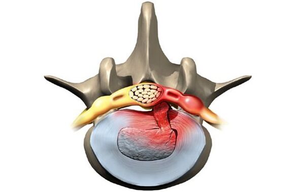
However, osteonecrosis in overwhelming cases affects the spine. Why so? The fact is that between the vertebrae there is a type of cushion - intervertebral discs (discs). The physiological role of these discs is to cushion and protect the vertebral bodies from premature wear from mechanical impact. The intervertebral disc consists of a loose inner nucleus surrounded by an arcuate fiber and an upper and lower cushion.
The disc is subjected to tremendous mechanical stress, resulting in permanent damage to its structures at the cellular level. In humans, these processes are all too obvious - this is our payment for vertical walking. To prevent the disk from being completely "erased", it must be continuously regenerated, that is, restored. It is the balance of the damage-regenerating processes that determines the normal structure of the disc. Another curious detail is that the blood and nutrient supply to the intervertebral discs is not accomplished through the overgrown blood vessels in childhood but diffuses from the bony tissue of the vertebral bodies. Again, the payment for mobility is two limbs, not four.
As a result, the intervertebral discs are anatomically and physiologically vulnerable. Any negative process in the body leads to an imbalance in the repair of damage and leads to the development of dystrophy and degeneration in the disc. A structurally defective disc can no longer withstand proper mechanical stress. Under excessive pressure from the upper vertebrae, the discs are displaced in different directions, usually laterally and posteriorly. This process is called a herniated disc.
The bony tissue of the vertebrae that has lost its cartilage lining is also subjected to mechanical wear and tear. Due to constant trauma to the anterior edge surface of the vertebral bodies, pathological foci of bone are formed - bone tumors. Spondylolisthesis develops. Due to the degeneration and displacement of the discs, the disc space is reduced, the spinal canal narrows, and the roots of the spinal nerves are compromised. borehole.
Reason
The causes, or etiological factors, of osteonecrosis are diverse. They can be both local, i. e. caused by pathology of the spine itself and general disorders at the organ level. Any pathology that leads to a violation of the structure of the spine or a metabolic disorder can be considered the cause of osteonecrosis. In this regard, there are:
- Changes in the configuration of the spine (scoliosis, scoliosis, or scoliosis)
- Other musculoskeletal defects - flat feet, narrow shoulders, pelvic deformities
- Spinal cord injury
- Weak immunity
- Metabolic disorders - osteoporosis, obesity, diabetes, thyroid disease
- Diseases of the cardiovascular system - atherosclerosis, hypertension
- Digestive disorders that lead to insufficient absorption of nutrients from the digestive tract
- Genetic.
It should be noted that the above medical conditions do not necessarily lead to osteonecrosis. This requires constant exposure to several risk factors - hypothermia, malnutrition, a sedentary lifestyle, or conversely, excessive physical exertion.
Symptom
Osteochondrosis is itself an asymptomatic process. And at the same time, signs of disc degeneration are also very diverse. How so? The fact is that the clinical manifestations of osteonecrosis are based on its complications - herniated disc, spondylolisthesis, sciatica, spinal stenosis.
Furthermore, the clinic is highly variable depending on the predominant location of the process in the cervical, thoracic or dorsal spine. The last part is most often affected, as the lower back is where physical activity is maximal. Signs of light zone osteonecrosis:
- Pain (paralysis, low back pain, sciatica)
- Limited movement of the lower back and lower extremities (intermittent movements)
- Here, sensitization disorder of the paresthesia type - numbness, burning, shivering
- Lumbar muscle strain disease
- In the absence of treatment, dysfunction of the pelvic organs.
Cervical fibroids are observed less frequently than cervical fibroids. However, this disease is also quite common. In addition to typical signs such as pain (cervical pain), decreased sensitivity and movement in the upper extremities, tarsal osteosarcoma caused by impaired blood supply to the brain has its own characteristics. These features are demonstrated:
- Insomnia
- Headache, Dizziness
- Periodic nausea
- General weakness, rapid fatigue
- Blood pressure fluctuations
- Occasional toothache
- Behavioral reactions in the form of tears, irritability.
The affected area of the chest with osteonecrosis is relatively rare. The patients in this case are people who are forced by their profession to sit in an uncomfortable position - schoolchildren, students, programmers, office workers. Symptoms of osteonecrosis in this case will be as follows:
- Pain and paresthesia in the chest
- Shortness of breath
- Heartbeat feeling
- Limit movement of the thoracic spine.
Diagnose
From all this, it is clear that osteonecrosis is a chameleon disease. Because of the same signs, it is easy to confuse with cerebrovascular accident, hypertension, myocardial infarction, angina pectoris, autonomic nervous disorder. That is why, for an accurate diagnosis, a comprehensive, complex diagnosis is required to accurately identify the symptoms and treat the osteonecrosis.
This diagnosis, in addition to traditional questioning and clarifying patient complaints, should include a medical examination and special research methods. These methods include X-ray of the spine, ultrasound of internal organs. Recently, computers and magnetic resonance imaging have been successfully used to diagnose osteonecrosis.
The treatment
Therapeutic strategies for osteonecrosis include the use of:
- Drugs
- Massage
- Physiotherapy procedures
- Physical therapy (exercise therapy)
- Manual therapy
- Acupuncture.
Drugs for the treatment of osteonecrosis are mainly aimed at reducing pain and eliminating inflammatory processes in the nerve roots. For this purpose, NSAID drugs are used. In various combinations, these drugs are widely used in the form of ointments, injections, and tablets to treat osteonecrosis. It should not be forgotten that these drugs have an adverse effect on the liver, stomach and intestines. In this way, they can exacerbate the metabolic disturbances in osteonecrosis. They relieve pain due to good blockade with local anesthetics. Yes, the effects of these funds are short-lived, and have no effect on the overall process of bone loss.
It is possible to improve metabolism at the body and local level with the help of drugs such as chondroprotectors, immunostimulants and vitamins with minerals. Chondroprotectors are used in tablets, ointments and ampoules. Among the fortifiers, vitamin C, group B, in combination with minerals are used. In this regard, calcium preparations are preferred above all. Indeed, contrary to some misconceptions, the basis of osteonecrosis is not excess but only calcium deficiency.
After successful exacerbation, physical therapy, massage, and exercise are shown. Calcium electrophoresis, hydrocortisone electrophoresis, amplipulse, paraffin therapy were used as physical procedures. All these measures are aimed at eliminating pain and inflammation in the nerve roots, ligaments and muscles. Therapeutic massage of osteonecrosis is performed according to a generally accepted method. The massage area is selected depending on the location of the osteonecrosis. The extension of range of motion is achieved with the help of exercise therapy. At the beginning, during the exacerbation phase, there is practically no dynamic load. The patient is continuously in an optimal position. At this time, it is desirable to wear immobilizers - a corset, a Shants' collar. Once the exacerbation is eliminated, the volume and duration of movements in exercise therapy are increased.
Recently, in the treatment of osteonecrosis, non-traditional methods of treatment have been applied - acupuncture, manual therapy, osteopathy. Acupuncture is the impact on specific biologically active points along the spine, on the backs of hands and feet. With manual therapy, the normal position of the vertebrae and discs is restored through the manual action of the therapist's hand. And during osteopathy, the structural integrity of the musculoskeletal system is ensured by specific techniques. In case there is no effect of conservative measures in the treatment of bone necrosis, persistent pain, complications, surgery is indicated. Pathologically displaced discs are removed. Currently, for this purpose, a microdiscectomy is performed - endoscopic removal of a displaced disc.