


Coxarthrosis affects the hip joints of middle-aged and elderly people. The causes of its development are previous injuries, congenital and acquired diseases of an inflammatory or non-inflammatory nature. The leading symptoms of coxarthrosis are hip pain, morning swelling, and stiffness with movement. In the early stages of the pathology, treatment is conservative. If it is ineffective in the setting of rapidly progressing coxarthrosis or detected late, surgical intervention, usually endoscopic, is indicated.
Pathology description
Coxarthrosis (osteoarthrosis, arthrosis deformans) is a degenerative-dystrophic disease of the hip joint. At the initial stages of development, the structure of synovial fluid changes. It becomes viscous, thick and therefore loses the ability to nourish hyaline cartilage. Due to dehydration, its surface dries out and becomes covered with many radial cracks. In this condition, the hyaline cartilage does not soften well the shocks when the bones forming joints come into contact.
To adapt to the increasing pressure on them, bone structures are deformed with the formation of growth cells (osteoporosis). Metabolic processes in the hip joint are impaired, negatively affecting the muscles and ligamentous apparatus of the joint.
Degree
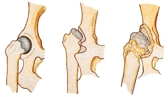
Each stage is characterized by its own symptoms, the severity of which depends on the degree of narrowing of the joint space and the amount of bone growth formed.
| Severity of coxarthrosis | Characteristic symptoms and radiological signs |
|---|---|
| Firstly | The joint space was unevenly narrowed and single bone spurs had formed around the acetabulum. There is a slight feeling of discomfort but the disease usually does not manifest clinically |
| Monday | The joint space is nearly 2 times narrower, the femoral head is misaligned, deformed, enlarged and the bone grows even outside the cartilage lip. Hip pain becomes constant and is accompanied by significant limitation of mobility |
| The third day | Complete or partial fusion of the joint space, multiple bone growth, widening of the femoral head. The pain occurs day and night and spreads to the thighs and legs. Movement is possible only with the help of a cane or crutches |
Cause of the disease
Primary coxarthrosis is a destructive degenerative lesion of the hip joint, the cause of which is unknown. This means that no prerequisites for premature destruction of hyaline cartilage have been identified. The following medical conditions can cause secondary coxarthrosis:
- previous injury - fracture of the femoral neck or pelvis, dislocation;
- hip dysplasia;
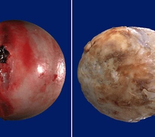
- aseptic necrosis of the femoral head;
- congenital hip dislocation;
- inflammation, including infectious diseases of the joints (rheumatism, reactive arthritis, gout, tendinitis, bursitis, synovitis).
Prerequisites for the development of coxarthrosis are obesity, increased physical activity, sedentary lifestyle, metabolic disorders, hormonal disorders, kyphosis, scoliosis and flat feet.
Symptoms of the disease
At the initial stages of development, coxarthrosis can manifest itself only with mild pain. They often occur after intense physical exertion or a hard day at work. This person attributed the deterioration to muscle "fatigue" and did not seek medical help. This explains the frequent diagnosis of coxarthrosis in stages 2 or 3, when conservative therapy is ineffective.
Restriction of joint mobility
Range of motion in the hip joint is reduced due to compensatory growth of bone tissue, damage to the synovial membrane, and replacement of areas of the joint capsule with fibrous tissue that does not have any functional activity. Mobility may be somewhat limited even with grade 1 coxarthrosis. Difficulty arises when performing rotational movements with the legs.
As the disease progresses, morning stiffness and joint swelling become common. To regain mobility, a person must warm up for a few minutes. By lunchtime, range of motion is restored, including the production of hormone-like substances in the body.
crispy
When walking, bending and (or) extending the hip joint, clicking, crunching and clicking sounds can be clearly heard. The reason for the sound that accompanies each step is due to the friction of bone surfaces, including bone cells, against each other. Crepitus can also occur in normal health due to the rupture of carbon dioxide bubbles in the joint cavity. Coxarthrosis is manifested by its combination with dull or sharp pain.
Pain
The pain becomes constant in stage 2 of coxarthrosis. Their severity lessens somewhat after a long period of rest. Pain increases with subsequent recurrence or the development of synovitis (synovitis) that often accompanies osteoarthritis. During the remission period, the feeling of discomfort decreases somewhat. But as soon as a person becomes hypothermic or lifts a heavy object, severe pain appears again.
Muscle spasms
Increased tension in the skeletal muscles of the thigh occurs with coxarthrosis for a number of reasons. First, the ligaments weaken. The muscles contract to hold the head of the femur in the acetabulum. Second, increased tone often accompanies synovitis. Third, when bone spurs are displaced, nerve endings are compressed and muscle spasm becomes a compensatory response to acute pain.
lameness
In the later stages of coxarthrosis, the patient begins to limp. Changes in gait are caused by shrinkage and deformation of the bone surfaces, making it impossible for the legs to maintain an upright position. The person also limps to reduce the severity of the pain by transferring body weight to the unaffected limb.
Shorten the legs
Short legs of 1 cm or more are typical of grade 3 coxarthrosis. The cause of reduced length of the lower limbs is severe muscle atrophy, thinning and collapsing cartilage, narrowing of the joint space and deformity of the femoral head.
Diagnostic method
The initial diagnosis is made based on the patient's complaints, external examination, medical history, and results of several functional tests. Many inflammatory and non-inflammatory pathologies are disguised as symptoms of coxarthrosis, so instrumental and biochemical studies are performed.
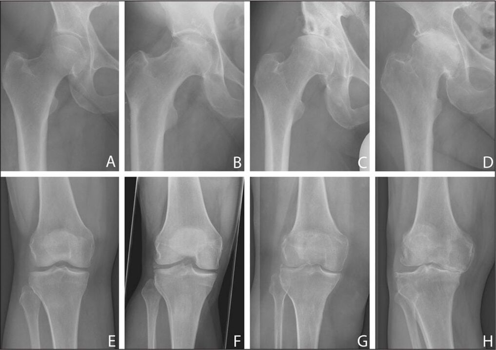
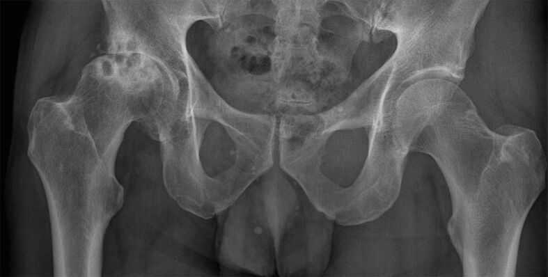
X-ray exam
The stage of coxarthrosis is determined by performing an X-ray examination. The images obtained clearly show destructive changes in the hip joint. This is a condition of narrowing joint space, deforming bone surfaces and forming bone spurs.
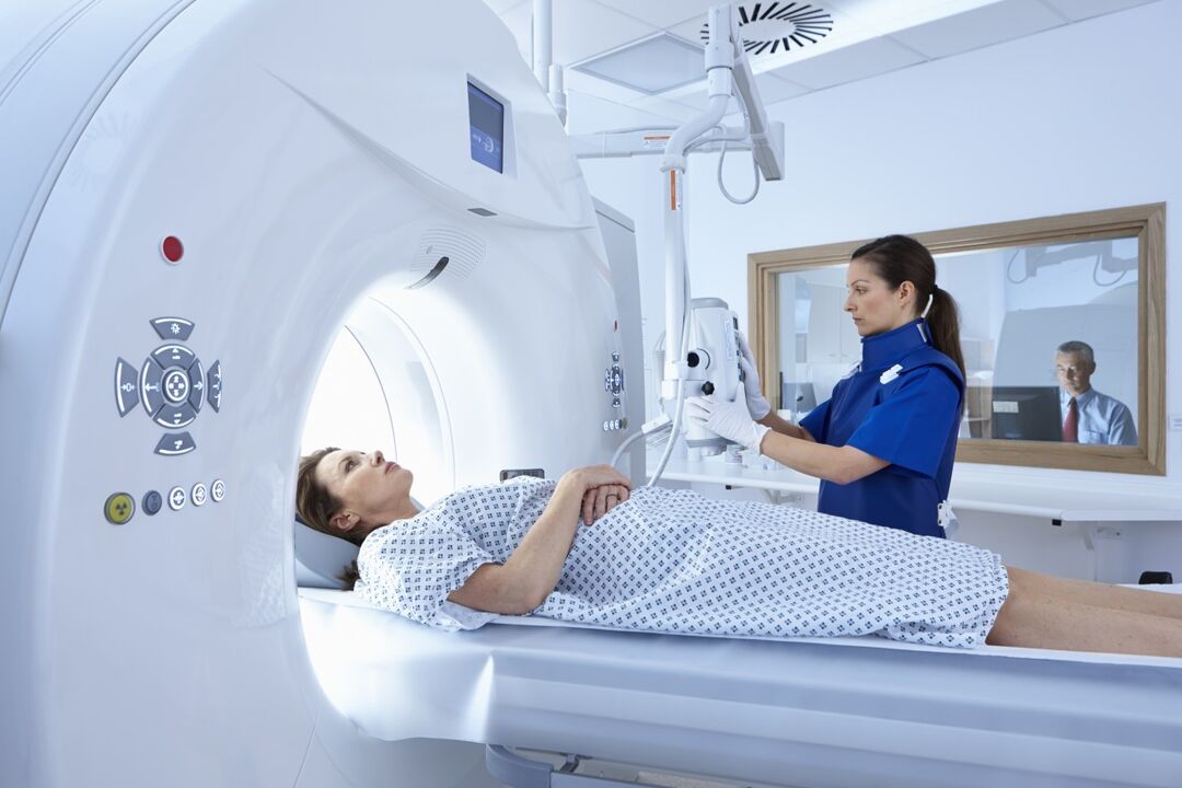
CT scan
CT scan is indicated for the patient to determine the degree of collapse and deformation of the hyaline cartilage. Research results also help evaluate the condition of the ligament-tendon apparatus, nerve trunks, muscles, small and large blood vessels.
Magnetic resonance imaging
MRI is one of the most informative studies in the diagnosis of coxarthrosis. To identify circulatory disorders in the affected joint area, it is performed with contrast. A periodic study is indicated to determine the degree of ligament damage and deformity of the femoral head, and to detect areas of fibrous degeneration of the joint capsule.
Measure foot length
Before measuring, the doctor asks the patient to stand up and straighten their legs as much as possible. To obtain the most reliable data, orthopedists use two bony landmarks. Superior - the anterior axis of the pelvis, located on the anterolateral surface of the abdomen at the outer edge of the inguinal ligament. The second reference point is any bony structure of the knee, ankle or heel. Measuring leg length may not be very informative if coxarthrosis affects two hip joints at the same time.
Laboratory research
Clinical blood and urine tests are performed to evaluate the patient's general health. And the results of biochemical studies often help to detect pathologies that caused the development of coxarthrosis. Gouty arthritis is manifested by high levels of uric acid and its salts. An increase in the erythrocyte sedimentation rate and an increase in the number of leukocytes indicate the presence of an inflammatory process (synovitis, arthritis, synovitis). To rule out rheumatoid arthritis, rheumatoid factor, C-reactive protein, and antinuclear antibodies are determined.

Puncture the hip
Using a puncture, synovial fluid is collected to study its composition and detect changes in consistency. If an inflammatory process is suspected, further biochemical examination of the biological sample should be performed.
Treatment options
When determining treatment tactics, the orthopedist will take into account the severity of coxarthrosis, the form of its course, the causes of its development and the severity of symptoms. Patients are often advised to wear bandages with stiff ribs and orthotics from the first days of treatment. The use of orthopedic devices helps slow down the process of cartilage breakdown and bone deformation.
Medicines
In the treatment of deforming arthropathy, drugs of various clinical and pharmacological groups are used. These are nonsteroidal anti-inflammatory drugs (NSAIDs), muscle relaxants, glucocorticosteroids, chondroprotectors, ointments and gels with a warming effect.
lockdown
To relieve acute pain that cannot be eliminated by NSAIDs, intra-articular or peri-articular blockers are prescribed. To carry them out, hormonal agents are used. The analgesic effect of glucocorticosteroids is enhanced when combined with anesthetics.
Injections
Intramuscular injection of NSAID solutions allows you to eliminate severe pain in the hip joint. To relax skeletal muscles, people often use a drug that, in addition to muscle relaxants, also includes anesthetics. In injectable form, the treatment regimen includes B vitamins, drugs that improve blood circulation, and drugs that protect cartilage.
Diet therapy
Overweight patients are advised to lose weight to slow the spread of the disease to healthy joint structures. The calorie content of the daily menu should be limited to 2000 kilocalories by excluding foods rich in fats and simple carbohydrates. Nutritionists recommend that all patients with coxarthrosis adhere to proper nutrition. The diet should include fresh vegetables, fruits, berries, cereals, fatty sea fish and dairy products. Following a therapeutic diet will stimulate the strengthening of the immune system and improve overall health.
Exercise therapy and massage
In the treatment of coxarthrosis, classical massage, acupressure and vacuum therapy are used. After several training sessions, blood circulation in the hip joint improves and nutrient reserves are replenished. Carrying out stimulating massage procedures strengthens the ligament-tendon apparatus and restores soft tissues damaged by the displacement of bone cells.
Regular exercise therapy is one of the most effective ways to treat osteoarthritis. A set of exercises is compiled specifically for the patient by a physiotherapist, taking into account the patient's physical strength.
Physical therapy
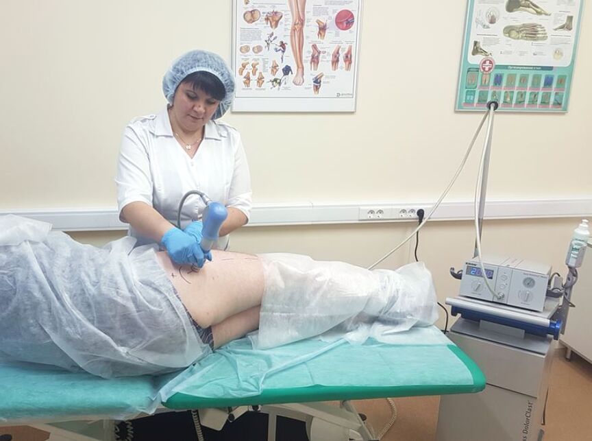
Patients with coxarthrosis are prescribed up to 10 sessions of magnetic therapy, laser therapy, UHF therapy, ultraviolet irradiation and shock wave therapy. The therapeutic effect of the procedures is due to improved blood circulation, acceleration of metabolism and regeneration. To relieve acute pain, electrophoresis or ultrasound is performed with glucocorticosteroids, anesthetics and B vitamins. Application with ozokerite or paraffin eliminates discomfort.
Surgical intervention
If conservative treatment is ineffective, the pain cannot be eliminated with medication or there is stable progression of coxarthrosis, the patient should undergo surgical intervention. Surgery is performed immediately in cases of pathology of the 3rd degree of severity, since it is not possible to eliminate destructive changes in cartilage and bone using medications or exercise therapy.
joint surgery
The operation is performed using general anesthesia. The femoral head is removed from the acetabulum. Visible destructive changes in the tissue are corrected - bony growths are removed, the joint surface is leveled, necrotic tissue is excised. During surgery, cavities will be formed and filled with ceramic implants.
Endoscopic
Hip replacement with implants is performed under general anesthesia. To prevent the development of the infectious process, a course of antibiotic treatment is prescribed. After 10 days, the sutures were removed and the patient was discharged from the hospital. In the rehabilitation phase, patients are guided through physical therapy and massage and exercise therapy.
Possible consequences
At the final stage of the pathology, contractures and contractures develop. The patient's legs are constantly bent so he must use a cane or crutches to move. Once the joint is completely fused, the patient will be immobile, unable to do housework, and disabled. Coxarthrosis is often complicated by aseptic necrosis of the femoral head, knee osteoarthritis, and arthritis.
Prevention and prognosis
Only grade 1 coxarthrosis responds well to conservative treatment. In other cases, arthroscopy allows you to completely restore the functional activity of the hip joint. After installation of the prosthesis, the patient quickly returns to an active lifestyle.
To prevent the disease, orthopedic doctors recommend quitting smoking, abusing alcoholic beverages, doing daily physical therapy and exercise, and losing weight if necessary.